
Anemia: Symptoms, Causes, Iron Deficiency, Treatment, Foods & Pregnancy Guide
What is Anemia?
Anemia is a blood disorder where oxygen-carrying capacity drops due to a decrease in:
- 🔻 Red Blood Cells (RBCs)
- 🔻 Hemoglobin (Hb)
- 🔻 Packed Cell Volume (PCV / Hematocrit)
Result: Less oxygen reaches body tissues → fatigue, weakness
Classification of Anaemia
Anaemia is classified into two main types:
1. Morphological classification
2. Etiological classification.
1. Morphological Classification of Anemia
Based on RBC size and colour
RBC size → Determined by Mean Corpuscular Volume (MCV)
RBC colour → Determined by Mean Corpuscular Haemoglobin Concentration (MCHC) – Blood Test
| Type of Anaemia | Size of RBC (MCV) | Colour of RBC (MCHC- mchc blood test) |
| Normocytic Normochromic Anaemia | Normal | Normal |
| Normocytic Hypochromic Anaemia | Normal | Less |
| Macrocytic Hypochromic Anaemia | Large | Less |
| Microcytic Hypochromic Anaemia | Small | Less |

Visual guide to anaemia classification based on red blood cell size (MCV) and haemoglobin concentration (MCHC)
2. Etiological classification of Anemia
- Hemorrhagic anemia
- Hemolytic anemia
- Nutrition deficiency anemia
- Aplastic anemia
- Anemia of chronic diseases
Hemorrhagic anemia
- Haemorrhagic anaemia is anaemia caused by excessive blood loss (haemorrhage).
What Happens in Acute Hemorrhagic anemia?
- Sudden, severe blood loss (e.g.Accident)
- Plasma volume is replaced within ~24 hours
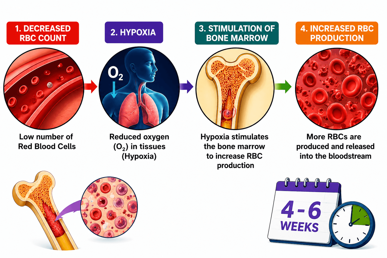
- RBC replacement takes 4–6 weeks
- Temporary haemodilution occurs due to fewer RBCs
- RBCs remain normocytic and normochromic
- Reduced RBC count → Hypoxia
- Hypoxia stimulates bone marrow to increase RBC production
- Anaemia usually corrects within 4–6 weeks
- Morphology: Normocytic Normochromic Anaemia

A step-by-step infographic showing how a decreased red blood cell (RBC) count causes hypoxia, stimulates the bone marrow, and increases red blood cell production, with recovery typically occurring within 4–6 weeks.
Chronic hemorrhage refers to the slow and continuous loss of blood over a prolonged period due to internal or external bleeding. Unlike acute hemorrhage, where blood loss occurs suddenly, chronic hemorrhage develops gradually and may remain unnoticed for a long time.
Common causes of chronic hemorrhage include:
- Peptic ulcers causing slow gastrointestinal bleeding
- Purpura leading to abnormal bleeding tendencies
- Hemophilia due to impaired blood clotting
- Menorrhagia causing excessive menstrual blood loss
How Chronic Hemorrhage Causes Anemia
Continuous blood loss results in the gradual depletion of the body’s iron stores because iron is an essential component of hemoglobin synthesis. As iron levels decrease, the bone marrow cannot produce enough hemoglobin for red blood cells (RBCs),
Chronic blood loss ➡️ Loss of iron from the body ➡️ Iron deficiency ⬇️ ➡️ Reduced hemoglobin synthesis ➡️ Formation of abnormal microcytic & hypochromic RBCs
Effect on Red Blood Cells
Due to reduced hemoglobin production, RBCs contain less hemoglobin and appear smaller than normal. These changes are characteristic of iron deficiency anemia:
- Microcytic RBCs → Red blood cells become smaller in size due to impaired hemoglobin production.
- Hypochromic RBCs → RBCs appear paler because they contain less hemoglobin.
Therefore, chronic hemorrhage commonly results in microcytic hypochromic anemia, where the reduced hemoglobin content affects the oxygen-carrying capacity of blood.
What is Hemolytic Anemia: Causes, Types, and Mechanism of RBC Destruction
Hemolysis refers to the destruction of red blood cells (RBCs) before their normal lifespan is completed. When excessive RBC destruction occurs and the bone marrow is unable to compensate by increasing RBC production, the condition is known as hemolytic anemia.
Normally, the bone marrow continuously produces new RBCs to replace old or damaged cells. However, in hemolytic anemia, the rate of RBC destruction exceeds the body’s ability to replace them, leading to a reduction in circulating RBCs and decreased oxygen-carrying capacity of blood.
Types of Hemolytic Anemia
Hemolytic anemia is broadly classified into two types:
- Extrinsic Hemolytic Anemia
- Intrinsic Hemolytic Anemia
Extrinsic Hemolytic Anemia
Extrinsic hemolytic anemia occurs when healthy RBCs are destroyed due to external factors acting outside the blood cells. These factors damage normal RBCs and increase their breakdown, resulting in excessive hemolysis.
When RBC destruction is caused by immune-related mechanisms, especially due to antibodies attacking RBCs, it is also known as autoimmune hemolytic anemia.
What is the most common cause of hemolytic anemia?
Mechanism of Extrinsic Hemolytic Anemia
External factors
➡️ Destruction of healthy RBCs
➡️ Increased hemolysis
➡️ Reduced RBC lifespan
➡️ Bone marrow unable to compensate
➡️ Development of hemolytic anemiaWhat is the most common cause of Causes of Extrinsic Hemolytic Anemia
Several conditions, infections, medications, and chemicals can trigger extrinsic RBC destruction.
1. Medical Conditions
- Liver failure – Impaired liver function can contribute to abnormal RBC breakdown.
- Renal disorders – Kidney-related abnormalities may affect RBC survival.
- Hypersplenism – Excessive activity of the spleen causes increased removal of RBCs.
- Severe burns – Damage to RBC membranes can lead to hemolysis.
2. Infections
Certain infections can increase RBC destruction, including:
- Hepatitis infections
- Malaria
- Septicemia (bloodstream infection)
3. Drug-Induced Hemolysis
Some medications can trigger immune reactions or directly damage RBCs, including:
- Penicillin
- Antimalarial drugs
- Sulfa drugs
4. Chemical Poisoning
Exposure to certain toxic substances may damage RBCs and cause hemolysis, including:
- Lead poisoning
- Coal and tar-related chemicals
5. Immune-Mediated Causes
The immune system may mistakenly attack RBCs, leading to their destruction. Examples include:
- Isoagglutinins such as anti-Rh antibodies
- Autoimmune diseases, including:
- Rheumatoid arthritis
- Ulcerative colitis
Intrinsic Hemolytic Anemia: Causes, Mechanism, and Examples
Intrinsic hemolytic anemia occurs when red blood cells (RBCs) are destroyed due to inherent defects within the RBCs themselves. In this condition, the body produces abnormal or unhealthy RBCs that have a shorter lifespan and are destroyed earlier than normal.
Unlike extrinsic hemolytic anemia, where external factors damage healthy RBCs, intrinsic hemolytic anemia results from problems that are present inside the RBCs. These defects may affect the shape, structure, or function of RBCs, making them more fragile and easily broken down.
Mechanism of Intrinsic Hemolytic Anemia
Defective RBC production
➡️ Formation of abnormal and fragile RBCs
➡️ Reduced RBC lifespan
➡️ Increased destruction of RBCs (hemolysis)
➡️ Development of hemolytic anemiaIntrinsic hemolytic anemia is commonly inherited and is associated with genetic abnormalities affecting RBC structure and hemoglobin production.
Common Examples of Intrinsic Hemolytic Anemia
1. Sickle Cell Anemia
Sickle Cell Anemia
Sickle cell anemia is an inherited blood disorder in which red blood cells (RBCs) become abnormally sickle- or crescent-shaped. It is caused by a specific form of sickle cell disease known as hemoglobin SS (HbSS) disease and is more common in people with ancestry from regions where malaria has historically been common, including parts of Africa.
What Causes Sickle Cell Anemia?
Sickle cell anemia is caused by an abnormal form of hemoglobin called hemoglobin S (HbS).
In hemoglobin S:
- The alpha (α) globin chains are normal.
- The beta (β) globin chains contain an abnormality.
- Under low-oxygen conditions, HbS molecules can polymerize into long, rigid fibers inside RBCs.
- This changes normally flexible, disc-shaped RBCs into rigid sickle-shaped cells.
These abnormal RBCs are fragile and break down more easily, causing hemolysis (destruction of red blood cells) and chronic hemolytic anemia.
How Is Sickle Cell Anemia Inherited?
Sickle cell anemia is an autosomal recessive genetic disorder. HbSS disease occurs when a person inherits two sickle cell (HbS) genes — one from each parent.
A person who inherits only one HbS gene usually has sickle cell trait rather than sickle cell anemia.
Why Do Sickle Cells Block Blood Vessels?
Sickled RBCs are less flexible than normal red blood cells. They can become trapped in small blood vessels and obstruct blood flow, particularly during sickling episodes.
This is known as vaso-occlusion and can reduce oxygen delivery to tissues, causing pain and tissue ischemia or infarction.
Sickle Cell Anemia in Children
Children with sickle cell disease may develop dactylitis (hand-foot syndrome) when vaso-occlusion affects the small blood vessels supplying the bones of the hands and feet.
This can cause:
- Painful swelling of the hands and feet
- Bone and tissue ischemia
- Recurrent pain episodes
Because sickled RBCs are destroyed more rapidly than normal RBCs, increased hemoglobin breakdown can also raise bilirubin levels, contributing to jaundice.
Quick Summary
HbS mutation → HbS polymerization → RBC sickling → increased RBC fragility → hemolysis → anemia
At the same time:
Sickled RBCs → vaso-occlusion → reduced blood flow → ischemia, pain and organ complications
2. Thalassemia
Thalassemia
Thalassemia is an inherited blood disorder in which the body does not produce normal amounts of the alpha (α) or beta (β) globin chains needed to make hemoglobin.
This imbalance reduces effective red blood cell production and causes RBCs to break down prematurely, resulting in hemolytic anemia.
Thalassemia is particularly common among people with ancestry from the Mediterranean, Middle East, South Asia and Southeast Asia.
What Causes Thalassemia?
Normal adult hemoglobin contains balanced amounts of α-globin and β-globin chains.
In thalassemia, genetic variants reduce or prevent the production of one type of globin chain. The remaining chains are produced in excess and can accumulate within developing RBCs.
This causes:
- Ineffective erythropoiesis – RBCs are not produced effectively in the bone marrow.
- Hemolysis – abnormal RBCs are destroyed prematurely.
- Anemia – reduced numbers of healthy circulating RBCs.
There are two main types:
1. Alpha (α) thalassemia
2. Beta (β) thalassemiaAlpha (α) Thalassemia
In alpha thalassemia, production of α-globin chains is reduced or absent.
The severity depends largely on how many of the four α-globin genes are affected.
Reduced α-chain production causes excess:
γ-globin chains in fetal life → Hb Bart’s (γ₄)
β-globin chains after birth → HbH (β₄)
These abnormal hemoglobins contribute to ineffective erythropoiesis and hemolysis.
The most severe form, alpha thalassemia major (Hb Bart’s hydrops fetalis), can cause severe fetal anemia and hydrops fetalis and may be fatal without specialist treatment.
Beta (β) Thalassemia
In beta thalassemia, production of β-globin chains is reduced or absent.
This leaves excess α-globin chains, which accumulate and precipitate within developing red blood cells.
The result is:
Reduced β-globin synthesis → excess α-chains → RBC damage → ineffective erythropoiesis + hemolysis → anemia
The clinical severity varies considerably, ranging from beta thalassemia trait to more severe forms requiring specialist management.
Alpha vs Beta Thalassemia – Quick Difference
Feature Alpha Thalassemia Beta Thalassemia Main defect ↓ α-globin production ↓ β-globin production Excess chains γ chains in fetus; β chains after birth α chains RBC effect Ineffective erythropoiesis + hemolysis Ineffective erythropoiesis + hemolysis Result Anemia Anemia Quick Summary
Thalassemia → defective globin-chain production → globin-chain imbalance → RBC damage → ineffective erythropoiesis + hemolysis → anemia
Key point: Unlike sickle cell anemia, where abnormal HbS causes RBCs to become sickle-shaped, thalassemia is primarily a problem of reduced globin-chain production.
Key Point
In both sickle cell anemia and thalassemia, abnormal RBC shape and structure increase their vulnerability to hemolysis, leading to a reduced number of circulating RBCs and the development of hemolytic anemia.
Intrinsic hemolytic anemia: abnormal red blood cells in sickle cell anemia and thalassemia become fragile and undergo hemolysis, contributing to anemia.
Nutritional Deficiency Anemia
Nutritional deficiency anemia occurs when the body does not get enough nutrients needed to produce healthy red blood cells (RBCs) and hemoglobin.
Important nutrients required for normal erythropoiesis (RBC production) include:
- Iron
- Vitamin B12
- Folate (vitamin B9)
- Protein
- Other nutrients that support normal blood formation, including vitamin C
Deficiency of these nutrients can reduce RBC production and lead to different types of anemia.
The main types of nutritional deficiency anemia are:
- Iron deficiency anemia
- Protein deficiency anemia
- Pernicious anemia (Addison’s anemia)
- Megaloblastic anemia
Iron Deficiency Anemia
Iron deficiency anemia is the most common type of anemia worldwide. It develops when the body does not have enough iron available to produce ade
quate amounts of hemoglobin.
Hemoglobin is the oxygen-carrying protein inside red blood cells. When iron is deficient, RBCs typically become:
Microcytic → smaller than normal
Hypochromic → contain less hemoglobin and appear paler
What Causes Iron Deficiency Anemia?
The major causes of iron deficiency anemia include:
- Blood loss – such as heavy menstrual bleeding or gastrointestinal blood loss.
- Low dietary iron intake – insufficient iron obtained from food.
- Reduced iron absorption – for example, due to gastrointestinal disorders or surgery affecting absorption.
- Increased iron requirements – particularly during pregnancy and periods of rapid growth.
Symptoms and Signs of Iron Deficiency Anemia
Common anemia symptoms can include tiredness, weakness, shortness of breath, dizziness, headaches and palpitations.
More characteristic features of iron deficiency may include:
- Brittle nails
- Koilonychia – thin, spoon-shaped nails
- Brittle or thinning hair
- Glossitis – a sore, smooth tongue caused by loss of normal papillae
- Angular cheilitis – soreness or cracking at the corners of the mouth
- Pica – craving or eating non-food substances
- Dysphagia – difficulty swallowing, which can occur in severe iron deficiency


Common signs and symptoms of iron deficiency anemia include brittle nails, koilonychia, thinning hair, glossitis, angular cheilitis, pica and, in severe cases, dysphagia.
Why Does Iron Deficiency Cause Microcytic Hypochromic Anemia?
Iron is essential for heme and hemoglobin synthesis.
When iron stores become depleted:
Iron deficiency → reduced hemoglobin synthesis → smaller, paler RBCs → reduced oxygen-carrying capacity → anemia
This produces the characteristic microcytic, hypochromic blood picture associated with iron deficiency anemia.
Quick Summary
Iron deficiency → ↓ hemoglobin synthesis → microcytic + hypochromic RBCs → reduced oxygen delivery → symptoms of anemia
Key point: When iron deficiency anemia is identified, it is important to determine why the iron deficiency developed, particularly whether there is an underlying source of blood loss.
Hemoglobin itself is made of:
Hemoglobin = Heme (contains iron) + Globin (protein chains)
The globin component consists mainly of α (alpha) and β (beta) globin chains in normal adult hemoglobin (HbA). These chains are built from amino acids obtained from dietary protein.
So the pathway is:
Severe protein deficiency → ↓ amino acid availability → impaired globin/protein synthesis + impaired erythropoiesis → ↓ hemoglobin/RBC production → anemia
One important correction: I would not state that protein deficiency anemia is characteristically “macrocytic and hypochromic.” Protein-energy malnutrition can cause anemia through several mechanisms, and RBC morphology can vary, especially because iron, folate, vitamin B12 and other deficiencies may coexist.
Which dietary proteins?
It is not about one special protein. Adequate high-quality dietary protein supplies the essential amino acids required for protein synthesis—for example:
Eggs • milk/dairy • fish • meat • pulses • lentils • beans • soy • nuts and seeds
Pernicious Anemia and Megaloblastic Anemia: Causes, Symptoms and Key Differences
Pernicious anemia and megaloblastic anemia are closely related conditions involving abnormal red blood cell (RBC) development. Vitamin B12 or folate deficiency can interfere with normal DNA synthesis, producing unusually large and immature red blood cells (Reticulocytes).
Understanding the difference is particularly important because vitamin B12 deficiency can cause neurological problems, whereas folate deficiency typically does not.
What Is Pernicious Anemia?
Pernicious anemia is an autoimmune condition that causes vitamin B12 deficiency because the body cannot absorb vitamin B12 normally.
It is sometimes referred to historically as Addison’s anemia.
Normally, parietal cells in the stomach produce intrinsic factor, a protein required for the absorption of vitamin B12 in the terminal ileum.
In pernicious anemia, autoimmune processes affect gastric parietal cells and/or intrinsic factor. This reduces intrinsic factor availability and consequently decreases vitamin B12 absorption.
How Does Pernicious Anemia Develop?
The process can be remembered as:
Autoimmune process → ↓ intrinsic factor → ↓ vitamin B12 absorption → impaired DNA synthesis → abnormal RBC maturation → megaloblastic, macrocytic anemia
Vitamin B12 is essential for normal DNA synthesis, red blood cell formation and neurological function.
When vitamin B12 is deficient, developing RBCs cannot mature normally. This results in megaloblastic changes and typically produces macrocytosis, meaning the circulating red blood cells are larger than normal.
Signs and Symptoms of Pernicious Anemia
People with pernicious anemia may develop general symptoms of anemia as well as characteristic features of vitamin B12 deficiency.
Common signs and symptoms include:
- Fatigue and weakness
- Pale skin
- Mild jaundice
- Red, sore or smooth tongue (glossitis)
- Shortness of breath
- Palpitations
Vitamin B12 deficiency can also affect the nervous system, causing:
- Paresthesia – numbness, tingling or pins and needles
- Muscle weakness
- Problems with balance
- Ataxia – impaired coordination
- Other neurological or cognitive changes in more severe or prolonged deficiency
Why Does Pernicious Anemia Cause Neurological Symptoms?
Vitamin B12 is required for normal nervous system function as well as blood formation.
Therefore:
Vitamin B12 deficiency → neurological dysfunction → numbness/tingling → weakness → balance and coordination problems
Neurological complications are particularly important because prolonged vitamin B12 deficiency can potentially cause irreversible neurological damage.
Pernicious Anemia and Autoimmune Disease
Pernicious anemia is more common in older adults and is associated with other autoimmune conditions.
These can include:
- Autoimmune thyroid disease
- Addison’s disease
- Other autoimmune disorders
This association occurs because pernicious anemia itself has an autoimmune basis.
What Is Megaloblastic Anemia?
Megaloblastic anemia is a type of macrocytic anemia caused by impaired DNA synthesis, most commonly due to deficiency of:
Vitamin B12 or folate (vitamin B9)
The developing blood cells continue to grow but cannot divide and mature normally. This produces abnormally large and immature precursor cells known as megaloblasts in the bone marrow.
The circulating red blood cells are typically macrocytic.
Folate Deficiency and Megaloblastic Anemia
Folate is essential for normal DNA synthesis and cell division.
When folate levels are inadequate:
Folate deficiency → impaired DNA synthesis → defective RBC maturation → megaloblastic changes → macrocytic anemia
Because red blood cells are continuously being produced, disruption of DNA synthesis can significantly affect normal erythropoiesis.
Symptoms of Folate Deficiency Megaloblastic Anemia
Possible features include:
- Fatigue
- Weakness

- Pale skin
- Shortness of breath
- Glossitis – red, sore or smooth tongue
- Other general symptoms of anemia

Vitamin B12 vs Folate Deficiency: The Key Difference
Both vitamin B12 deficiency and folate deficiency can cause megaloblastic macrocytic anemia, so their blood-related features can appear similar.
The major clinical distinction is neurological involvement:
| Feature | Vitamin B12 Deficiency | Folate Deficiency |
| Megaloblastic anemia | Yes | Yes |
| Macrocytosis | Yes | Yes |
| Impaired DNA synthesis | Yes | Yes |
| Glossitis | Can occur | Can occur |
| Neurological symptoms | Can occur | Typically absent |
| Paresthesia | Can occur | Usually absent |
| Ataxia/balance problems | Can occur | Usually absent |
Important Clinical Point
Folate should not simply be given to someone with an unexplained megaloblastic anemia without considering vitamin B12 deficiency.
Folate treatment can improve the anemia while an underlying vitamin B12 deficiency and its associated neurological complications remain untreated.
Quick Revision
Pernicious Anemia
Autoimmune disease → ↓ intrinsic factor → ↓ vitamin B12 absorption → B12 deficiency → impaired DNA synthesis → megaloblastic macrocytic anemia
Folate Deficiency
↓ Folate → impaired DNA synthesis → abnormal RBC maturation → megaloblastic macrocytic anemia
Aplastic Anemia: Causes, Blood Cell Changes and Key Features
Aplastic anemia is a rare but serious blood disorder in which the bone marrow fails to produce enough new blood cells.
Normally, red bone marrow contains hematopoietic stem cells, which produce:
- Red blood cells (RBCs) – carry oxygen
- White blood cells (WBCs) – help fight infection
- Platelets – help blood clot
In aplastic anemia, the bone marrow becomes hypocellular, with a marked reduction in blood-forming cells and increased replacement by fatty tissue.
This can result in pancytopenia, meaning reduced levels of RBCs, WBCs and platelets.
What Causes Aplastic Anemia?
In many people, no definite cause can be identified. This is known as idiopathic aplastic anemia.
In other cases, aplastic anemia may be associated with:
- Autoimmune destruction of hematopoietic stem cells
- Ionising radiation
- Certain medicines and toxic chemicals, including benzene
- Viral infections, particularly some forms of hepatitis
- Certain inherited bone marrow failure disorders
Some older sources list substances such as quinine, gold salts and radium. However, causes and drug associations should be assessed using current clinical evidence rather than assuming that every historical association is a common modern cause.
What Happens in Aplastic Anemia?
The basic mechanism is:
Bone marrow stem-cell damage → ↓ blood cell production → pancytopenia
This produces three important consequences:
↓ RBCs → anemia → fatigue, weakness, pallor and breathlessness
↓ WBCs → leukopenia/neutropenia → increased risk of infection
↓ Platelets → thrombocytopenia → easy bruising and bleeding
What Do the RBCs Look Like?
Aplastic anemia is generally considered a normocytic anemia, meaning the red blood cells are usually approximately normal in size.
The anemia results primarily from reduced production of RBCs by the bone marrow, rather than a primary defect in hemoglobin synthesis.
Key Point
Aplastic anemia is bone marrow failure, not simply an RBC disorder.
The easiest pathway to remember is:
Bone marrow failure → ↓ RBCs + ↓ WBCs + ↓ platelets → pancytopenia
Therefore, a patient may present with a combination of anemia, recurrent infections and abnormal bleeding or bruising.
Anemia of Chronic Disease: Causes, Mechanism and RBC Changes
Anemia of chronic disease (ACD), also called anemia of inflammation, is a common type of anemia associated with long-term inflammatory, infectious, autoimmune and malignant conditions.
Unlike iron deficiency anemia, the body may have adequate iron stores, but inflammation makes much of that iron less available for red blood cell (RBC) production.
What Causes Anemia of Chronic Disease?
During chronic inflammation, inflammatory cytokines increase production of hepcidin, a hormone mainly produced by the liver.
Hepcidin reduces the amount of iron absorbed from the intestine and limits the release of stored iron into the circulation.
The key pathway is:
Chronic inflammation → ↑ hepcidin → ↓ iron absorption & iron release → ↓ available iron → ↓ hemoglobin/RBC production → anemia
Other contributing mechanisms include:
- Reduced erythropoietin production or response
- Reduced bone marrow response to erythropoietin
- Shortened RBC lifespan
- Restricted availability of iron for erythropoiesis
Common Causes
Anemia of chronic disease may occur with:
- Chronic inflammatory diseases – e.g. rheumatoid arthritis
- Chronic infections – e.g. tuberculosis
- Chronic kidney disease (CKD)
- Cancer and other malignant conditions
Anemia in Chronic Kidney Disease
The kidneys produce erythropoietin (EPO), a hormone that stimulates the bone marrow to produce red blood cells.
In chronic kidney disease:
Kidney damage → ↓ erythropoietin → ↓ RBC production → anemia
Inflammation and altered iron metabolism can also contribute.
What Do the RBCs Look Like?
Anemia of chronic disease is typically:
Normocytic → RBCs are normal in size
Normochromic → RBCs have relatively normal coloration
However, particularly with prolonged disease, the anemia can sometimes become microcytic.
Anemia of Chronic Disease vs Iron Deficiency Anemia
Both conditions can involve reduced iron availability, but the underlying mechanisms differ.
Iron deficiency anemia: the body’s iron stores are genuinely depleted.
Anemia of chronic disease: iron may be present in storage, but inflammation restricts its availability for erythropoiesis.
Quick Revision
Chronic disease/inflammation → ↑ hepcidin → iron becomes less available → ↓ erythropoiesis → anemia
Key point: Think of anemia of chronic disease as “iron is present, but the bone marrow cannot access it efficiently.”
Reticulocyte: Definition, Normal Count and Clinical Significance
A reticulocyte is an immature red blood cell (RBC) that has recently been released from the bone marrow into the bloodstream.
Reticulocytes are slightly larger than mature red blood cells and contain small amounts of residual ribosomal RNA. This residual material forms a network-like appearance, called a reticulum, when stained with a supravital stain.
Why Is It Called a Reticulocyte?
The term reticulocyte comes from the fine reticular or network-like material seen inside the cell after supravital staining.
The pathway is:
Bone marrow → Reticulocyte → Mature RBC
As the reticulocyte matures, the remaining RNA and cellular material disappear, producing a fully mature red blood cell.
Reticulocyte Count
The reticulocyte count measures the percentage or number of reticulocytes circulating in the blood.
It is useful for assessing how actively the bone marrow is producing red blood cells.
A high reticulocyte count may indicate increased RBC production, for example after:
- Blood loss
- Hemolysis
- Successful treatment of certain anemias
A low reticulocyte count may suggest reduced or ineffective RBC production by the bone marrow.
Reticulocyte Count in Newborns
Newborn babies normally have a higher reticulocyte count than adults.
The reticulocyte count may be approximately:
2%–6% of circulating RBCs
This means around 2–6 reticulocytes per 100 red blood cells.
The level usually decreases after birth as red blood cell production adjusts to life outside the womb.
Why Is Reticulocyte Count Important in Anemia?
Reticulocyte count helps distinguish between anemia caused by increased RBC loss or destruction and anemia caused by reduced RBC production.
Anemia + high reticulocyte count → bone marrow is responding by increasing RBC production
Anemia + low reticulocyte count → bone marrow response may be inadequate
Leave a Reply