
Kidney Anatomy: Location, Structure, Function & Blood Supply
Kidney anatomy explains the location, structure, blood supply and function of the kidneys, two essential organs of the urinary system. Located in the posterior abdomen, the kidneys filter the blood, remove metabolic waste, regulate fluid and electrolyte balance, and contribute to blood pressure control and red blood cell production.
Understanding kidney anatomy provides an important foundation for learning about renal physiology, kidney diseases and overall kidney health.
Renal System Anatomy
The kidneys are a pair of retroperitoneal organs located on either side of the vertebral column. They play an essential role in maintaining fluid and electrolyte balance and removing waste products from the body.
In a healthy adult, each kidney extends approximately from the level of the 12th thoracic vertebra (T12) to the 3rd lumbar vertebra (L3). The right kidney is normally positioned slightly lower than the left kidney, mainly because of the space occupied by the liver.
Kidney Size and Weight
The size and weight of the kidneys vary between individuals and can also differ between males and females. In adults, a kidney is approximately:
- Length: 11–12 cm
- Width: 5.0–7.5 cm
- Thickness: 2.5–3.0 cm
The average kidney weight is approximately 125–170 g in adult males and 115–155 g in adult females.
Kidney size can also be assessed using magnetic resonance imaging (MRI). Normal kidney length and volume have a relatively wide range, reflecting differences in body size, sex and individual anatomy.

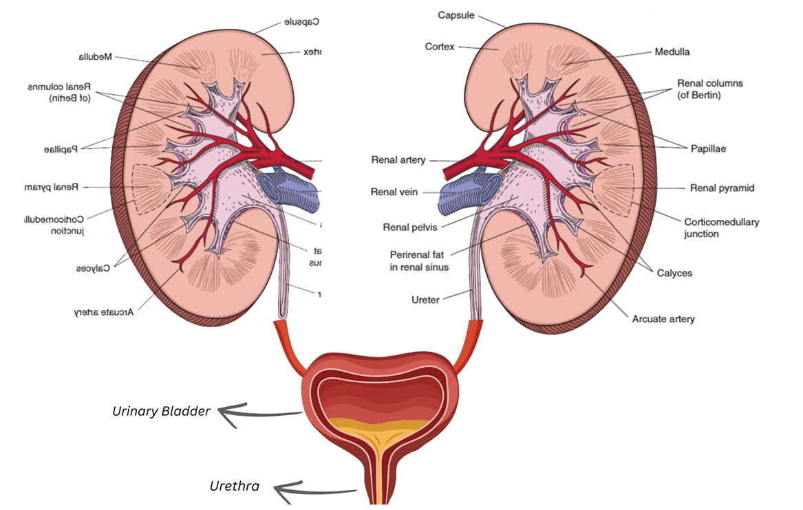
Kidney anatomy showing the retroperitoneal position, vertebral levels, relative size, and renal hilum of the right and left kidneys.
Renal Hilum
The renal hilum is a narrow opening located on the medial, or concave, border of each kidney. It provides a passage for important structures entering and leaving the kidney.
These structures include:
- Renal artery – supplies blood to the kidney
- Renal vein – carries blood away from the kidney
- Renal pelvis – collects urine before it passes into the ureter
- Lymphatic vessels
- Renal nerve plexus
These structures pass through the renal hilum and enter the renal sinus, an internal space within the kidney.
Fibrous Capsule of the Kidney
Each kidney is enclosed by a thin, tough fibrous capsule. Under normal conditions, the capsule has a smooth surface and can be separated relatively easily from the underlying kidney tissue.
Together, the position, size, external features and internal structures of the kidneys form an important part of normal renal anatomy and provide a foundation for understanding kidney function and renal disease.
Nephron Anatomy: Location, Structure and Role
The nephron is the structural and functional unit of the kidney. It is the microscopic structure responsible for filtering blood and helping the kidneys maintain the body’s fluid, electrolyte and acid–base balance.
Each kidney contains approximately 1 to 1.3 million nephrons. The number of functioning nephrons gradually decreases with age.
Structure of a Nephron
Each nephron has two main parts:
- Renal corpuscle
- Renal tubule
1. Renal Corpuscle
The renal corpuscle, also called the Malpighian corpuscle, forms the beginning of the nephron. It consists of the glomerulus, a network of capillaries, enclosed within Bowman’s capsule.
Blood is filtered across the glomerular filtration barrier, and the resulting filtrate enters Bowman’s capsule before passing into the renal tubule.

Renal corpuscle showing glomerulus and Bowman’s capsule involved in glomerular filtration
2. Renal Tubule
The renal tubule is the tubular portion of the nephron that begins at Bowman’s capsule. It consists mainly of the:
- Proximal convoluted tubule (PCT)
- Loop of Henle
- Distal convoluted tubule (DCT)
The filtrate passes through these segments, where water and useful substances are reabsorbed and selected substances are secreted.
The processed tubular fluid then enters the collecting duct, where further adjustment of water and electrolyte content occurs before the final urine passes towards the renal pelvis.
Anatomy of a Nephron

Anatomy of a nephron showing its structure and function, including the glomerulus, renal tubule and major sites of reabsorption involved in urine formation.

Nephron structure flowchart showing the major parts of the renal corpuscle and tubular portion, including the Loop of Henle and its thick and thin segments.
Main Parts of the Nephron Explained
Glomerulus
The glomerulus is a network of capillaries where filtration of the blood begins.
Bowman’s Capsule
Bowman’s capsule surrounds the glomerulus and collects the fluid filtered from the blood.
Proximal Convoluted Tubule (PCT)
The PCT is the major site of tubular reabsorption. It reabsorbs most of the filtered water and many useful substances, including glucose, amino acids, sodium and bicarbonate.
Loop of Henle
The loop of Henle helps establish the concentration gradient within the renal medulla that allows the kidneys to produce concentrated urine.
Distal Convoluted Tubule (DCT)
The DCT performs further selective reabsorption and secretion and contributes to the regulation of electrolytes and acid–base balance.
Collecting Duct
The collecting duct receives tubular fluid from multiple nephrons and carries it towards the renal pelvis. It also helps regulate the final concentration of urine, particularly under the influence of antidiuretic hormone (ADH).
Nephron Diagram: Labeled Structure and Parts

Nephron structure showing the renal corpuscle, proximal convoluted tubule, loop of Henle, distal convoluted tubule and collecting duct.
Nephron Structure and Function
| Nephron Part | Main Function |
| Glomerulus | Filters blood |
| Bowman’s capsule | Collects the filtrate |
| PCT | Major site of reabsorption |
| Loop of Henle | Creates the medullary concentration gradient |
| DCT | Fine-tunes electrolyte and acid–base balance |
| Collecting duct | Regulates final urine concentration and carries urine onwards |
Easy way to remember: Filter → Reabsorb → Concentrate → Fine-tune → Collect
Nephron Function: Filtration, Reabsorption, Secretion and Urine Formation
The nephron performs several processes that allow the kidneys to remove waste while maintaining the body’s fluid and electrolyte balance.

Functions of the nephron showing how filtration, reabsorption and tubular secretion help form urine and maintain fluid and electrolyte balance.
1. Glomerular Filtration
Blood is filtered at the glomerulus. Water and small molecules pass into Bowman’s capsule, while blood cells and most plasma proteins normally remain within the bloodstream.
2. Tubular Reabsorption
During tubular reabsorption, water and useful substances are transported from the tubular fluid back into the blood.
3. Tubular Secretion
During tubular secretion, selected substances are transported from the blood into the renal tubule. These include hydrogen ions, potassium ions and certain drugs and metabolites.
4. Urine Formation
After filtration, reabsorption and secretion, the remaining tubular fluid becomes urine. It passes through the collecting ducts towards the renal pelvis and then enters the ureter.
Where Does Reabsorption Occur in the Nephron?
Reabsorption occurs in several parts of the nephron, but the proximal convoluted tubule (PCT) is the major site of tubular reabsorption.
- PCT: Reabsorbs the majority of filtered water, sodium and many useful substances.
- Loop of Henle: The descending limb is highly permeable to water, while the ascending limb reabsorbs sodium, potassium and chloride and is relatively impermeable to water.
- DCT: Selectively reabsorbs electrolytes and contributes to electrolyte and acid–base regulation.
- Collecting duct: Regulates water reabsorption according to the body’s needs, particularly under the influence of ADH.
Key point: The PCT is the main site of tubular reabsorption.

Functions of the nephron showing how filtration, reabsorption and tubular secretion help form urine and maintain fluid and electrolyte balance.
Juxtaglomerular Apparatus: Structure and Function
The juxtaglomerular apparatus (JGA) is a specialised structure located at the vascular pole of each glomerulus, where the distal nephron comes into close contact with the afferent and efferent arterioles. It is an important part of kidney physiology because it helps control glomerular filtration rate (GFR), renal blood flow and blood pressure.
The juxtaglomerular apparatus has three main components:
- Macula densa
- Extraglomerular mesangial cells
- Juxtaglomerular (granular) cells


What is Macula Densa
The macula densa is a group of tightly packed specialised epithelial cells in the final part of the thick ascending limb, where it transitions into the distal convoluted tubule. It lies close to the afferent arteriole and senses the amount of sodium chloride (NaCl) reaching the distal nephron.
When NaCl delivery falls, the macula densa helps signal the juxtaglomerular cells to release renin. It also takes part in tubuloglomerular feedback, a local mechanism that adjusts arteriolar tone and helps stabilise renal blood flow and GFR.
Extraglomerular Mesangial Cells
Extraglomerular mesangial cells lie in the triangular region bordered by the afferent arteriole, efferent arteriole and macula densa. They are also called lacis cells, agranular cells or Goormaghtigh cells.
These cells form part of the JGA and are thought to help communication between the macula densa and the glomerular arterioles. They also produce signalling substances involved in local renal regulation.
Juxtaglomerular Cells
Juxtaglomerular cells, also known as granular cells, are modified smooth muscle cells found mainly in the wall of the afferent arteriole close to the glomerulus. Their cytoplasm contains secretory granules that store renin.
A concentration of these cells around the vascular pole has traditionally been described as the polar cushion or Polkissen.
Renin release increases mainly when:
- renal perfusion pressure falls;
- sympathetic stimulation increases; or
- sodium chloride delivery to the macula densa decreases.
Renin–Angiotensin–Aldosterone System (RAAS)
The renin–angiotensin–aldosterone system (RAAS) links the juxtaglomerular apparatus with the control of blood pressure, sodium balance and kidney function.

Renin–angiotensin–aldosterone system (RAAS) showing how renin and angiotensin II regulate blood pressure, GFR, sodium reabsorption, aldosterone and ADH.
Angiotensinogen → Renin → Angiotensin I → ACE → Angiotensin II → Aldosterone.
Renin released from JG cells converts liver-derived angiotensinogen into angiotensin I. Angiotensin-converting enzyme (ACE), found abundantly on vascular endothelium including the lungs, then converts angiotensin I into angiotensin II, the main active hormone of the system.
What Does Angiotensin II Do?
Angiotensin II has several important effects in renal system physiology:
- causes vasoconstriction, helping raise arterial blood pressure;
- preferentially constricts the efferent arteriole, helping maintain glomerular pressure and GFR when renal perfusion falls;
- stimulates aldosterone secretion from the adrenal cortex, increasing sodium retention;
- increases sodium reabsorption, particularly in the proximal tubule;
- stimulates thirst and ADH release, promoting water retention; and
- contracts glomerular mesangial cells, reducing the capillary surface area available for glomerular filtration.
Glomerular Mesangial Cells
Glomerular mesangial cells, also called intraglomerular mesangial cells, are located between the capillary loops inside the glomerulus. They form a supporting cellular network around the glomerular capillaries.
Their main functions include:
- providing structural support to glomerular capillaries;
- contracting to alter the surface area available for glomerular filtration;
- removing trapped particles and cellular debris by phagocytosis; and
- producing extracellular matrix and local signalling molecules such as cytokines and prostaglandins.
This contractile function means mesangial cells can influence GFR and therefore contribute to normal nephron function and filtration in the kidney.
Key difference: Extraglomerular mesangial cells are located outside the glomerular capillary tuft and form part of the JGA, while intraglomerular mesangial cells lie between the glomerular capillaries and directly support and regulate the filtration surface.
Juxtaglomerular Apparatus in Simple Terms
Think of the JGA as the kidney’s local pressure-and-salt sensor. The macula densa senses NaCl, the juxtaglomerular cells release renin, and the resulting RAAS response helps adjust blood pressure, sodium and water retention, renal blood flow and glomerular filtration rate.
Renal Circulation: Blood Flow Through the Kidney
Renal circulation is the movement of blood through the kidneys for glomerular filtration, tubular reabsorption and secretion. The kidneys receive roughly 20–25% of resting cardiac output, making renal blood flow unusually high for their size.
Renal Blood Flow and Renal Plasma Flow
Renal blood flow (RBF) is the total volume of blood delivered to the kidneys per minute, whereas renal plasma flow (RPF) refers only to the plasma component of that blood. RPF is therefore influenced by both renal blood flow and haematocrit. In renal physiology, effective renal plasma flow can be estimated using the clearance of substances such as para-aminohippurate (PAH).
Renal Blood Flow Pathway
Blood reaches the kidney through the renal artery, which arises from the abdominal aorta. The main pathway is:
Renal artery → Segmental arteries → Interlobar arteries → Arcuate arteries → Cortical radiate (interlobular) arteries → Afferent arteriole → Glomerular capillaries → Efferent arteriole → Peritubular capillaries or vasa recta → Renal veins
Segmental, Interlobar and Cortical Radiate Arteries
Inside the kidney, the renal artery divides into segmental arteries. These give rise to interlobar arteries, which travel between the renal pyramids. At the corticomedullary junction, they curve along the bases of the pyramids to form the arcuate arteries.
The arcuate arteries then give rise to cortical radiate arteries, traditionally called interlobular arteries. These extend into the renal cortex and give rise to numerous afferent arterioles supplying the glomeruli.
Afferent and Efferent Arteriole
The afferent arteriole carries blood into the glomerulus, where it divides into glomerular capillaries. These capillaries reunite to form the efferent arteriole, which carries blood away from the glomerulus.
This arrangement is important for GFR and glomerular filtration. Changes in resistance of the afferent and efferent arterioles alter glomerular capillary pressure and therefore influence filtration in the kidney.
Easy way to remember: Afferent arrives; efferent exits.
Peritubular Capillaries and Vasa Recta
After leaving the glomerulus, the efferent arteriole forms another capillary network. In cortical nephrons, this is mainly the peritubular capillary network, which surrounds the renal tubules and supports tubular reabsorption and secretion.
In juxtamedullary nephrons, efferent arterioles form the vasa recta. So, what is vasa recta? The vasa recta are long, straight capillaries that descend into the renal medulla alongside the loop of Henle and then return towards the cortex. They help preserve the medullary osmotic gradient needed to produce concentrated urine.
Where is vasa recta absent or highly reduced? It is essentially absent or only poorly developed in cortical nephrons, which have short loops of Henle. A well-developed vasa recta is a characteristic feature of juxtamedullary nephrons.
Renal Autoregulation and Tubuloglomerular Feedback
The kidneys can keep renal blood flow and GFR relatively stable across a range of arterial pressures through renal autoregulation. Two major mechanisms are involved:
- Myogenic response: Increased pressure stretches the afferent arteriole, causing its smooth muscle to contract. This limits excessive blood flow into the glomerulus.
- Tubuloglomerular feedback: The macula densa senses changes in NaCl delivery in the distal nephron and sends local signals that alter afferent arteriolar tone and renin release. This feedback helps stabilise renal blood flow and GFR.
Why Is Renal Circulation Important for Tubular Reabsorption?
The glomerular capillaries are specialised for filtration, whereas the relatively low-pressure peritubular capillaries favour reabsorption of water and solutes from the renal tubules back into the circulation. The vasa recta also helps maintain the medullary gradient required for water conservation.
The amount of water being reabsorbed in the collecting duct is controlled mainly by antidiuretic hormone (ADH, or vasopressin). ADH increases the water permeability of collecting-duct principal cells by promoting insertion of aquaporin-2 water channels. More ADH therefore promotes greater water reabsorption and produces a smaller volume of more concentrated urine; low ADH produces more dilute urine.
Renal Circulation: Key Points
- Segmental arteries are early branches of the renal artery within the kidney.
- Interlobar arteries travel between the renal pyramids.
- Arcuate arteries run along the corticomedullary junction.
- Cortical radiate arteries extend into the renal cortex and give rise to afferent arterioles.
- The afferent arteriole enters the glomerulus; the efferent arteriole leaves it.
- Peritubular capillaries mainly supply cortical nephron tubules.
- Vasa recta are associated with juxtamedullary nephrons and help maintain the medullary concentration gradient.
- Tubuloglomerular feedback helps regulate renal blood flow and GFR.
- ADH controls much of the variable water reabsorption in the collecting duct, helping determine whether urine becomes dilute or concentrated.
Kidney Functions
The kidneys do much more than produce urine. Their major functions include glomerular filtration, removal of metabolic waste, regulation of water and electrolytes, acid–base balance and long-term blood pressure control. They also have important endocrine functions.
One of these is the production of erythropoietin (EPO), mainly by specialised interstitial cells in the kidney. When renal oxygen availability falls, EPO production increases and stimulates the bone marrow to produce more red blood cells. The kidneys also activate vitamin D to calcitriol, which contributes to calcium and phosphate homeostasis.
Renal Function Tests
Renal function tests help assess how well the kidneys are filtering blood and maintaining normal biochemical balance. Common tests include serum creatinine, estimated glomerular filtration rate (eGFR), urea, electrolytes and urine albumin-to-creatinine ratio (ACR).
GFR is one of the most useful measures of kidney filtration. In routine clinical practice it is usually estimated as eGFR from serum creatinine together with factors used by the relevant estimating equation. Urine ACR provides additional information by detecting increased albumin loss in the urine.
Kidney Anatomy and Physiology: Key Takeaway
The nephron in the kidney links renal anatomy directly with renal system physiology. Blood reaches the glomerulus through the afferent arteriole, undergoes glomerular filtration, and leaves through the efferent arteriole. The renal tubules then modify the filtrate through tubular reabsorption and secretion, while the loop of Henle, vasa recta and collecting duct help determine urine concentration. At the same time, the macula densa, juxtaglomerular apparatus and tubuloglomerular feedback help regulate renal blood flow and GFR.
Leave a Reply